Platform that
Visualizes Prostate
Microvasculature

Super-Resolution Microvascular Imaging
Point of Care Diagnosis
Transforming the Prostate Pathway
Less Grey Imaging is a Med Tech company developing the next-generation ultrasound-based imaging software for prostate cancer diagnosis.
Â
Founded in 2023 out of Heriot-Watt University, Edinburgh, the founding team unites deep expertise in ultrasound physics, applied mathematics, machine learning, and clinical translation. Built on over a decade of foundational research, their technology is backed by 3 patent applications, unique clinical datasets, and published validation – engineered from the ground up to deliver clinically actionable imaging at the point of care.
Pioneering a new standard in prostate cancer diagnosis
Less Grey Imaging is redefining how prostate cancer is detected and diagnosed. Our proprietary platform, ProViu, transforms conventional ultrasound into high-resolution microvascular imaging — enabling clinicians to identify cancer earlier and make confident decisions during the patient visit.
The current diagnostic pathway is expensive, invasive, and inaccurate. Systematic biopsy misses more than half of clinically significant cancers, while MRI – the current gold standard – fails to detect up to 28% of cancers. Meanwhile, the majority of biopsies performed are unnecessary, exposing patients to procedural risk, infection, and significant anxiety.
ProViu addresses this gap by delivering MRI-comparable imaging performance using conventional ultrasound systems — without the infrastructure, delays, or constraints of traditional imaging workflows.
Core Technology
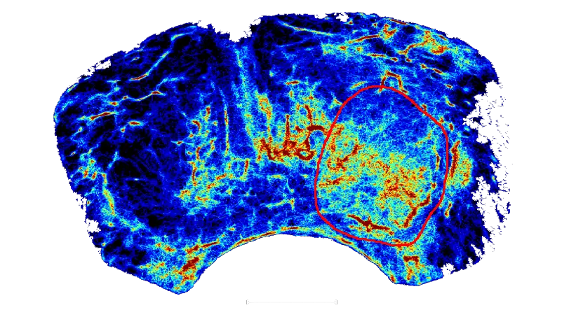
ProViu tracks microbubble contrast agents using conventional ultrasound systems to generate prostate maps of microvasculature. By overcoming the acoustic diffraction limit, the technology reveals vascular patterns associated with cancer that are invisible to conventional imaging and MRI.
Vessel Reconstruction
ProViu reconstructs microvascular structures at higher resolution than conventional ultrasound, restoring critical diagnostic detail and enabling earlier and more accurate clinically significant lesion detection.
Quantification of Microvasculature
Built-in assessment algorithms allow for the quantification of blood flow & speed, microbubble density as well as morphological vascular characteristics such as vessel diameter, length or their tortuosity. These metrics give clinicians an in–depth understanding of tumor vascularity – enabling them to detect, localize, and characterize suspicious lesions with greater precision, ultimately supporting more confident and accurate prostate cancer diagnosis.
Redefining imaging,
improving decisions